
Museum of human anatomy of ”Nicolae Testemitanu” University represents more than a simple display of exhibits, it is a unique collection, known at international level due to the number and quality of included specimens.
Senior assistant E.M Koblik-Zelter (1968-1989) directly contributed to the illustration design for most of the scientific theses and publications developed at the Department. The impeccable images of macro-, macromicro and microscopic anatomical pieces made by him put the finishing touches on the scientific papers of the Department’s academic staff and show the veracity of the obtained scientific results. Scientific investigations conducted by Professor B.Z. Perlin on the nervous system of conjunctival formations in normal, pathological and experimental conditions are widely known in the country and abroad, as many of them have been presented at national and international congresses.
At that time, the training of young lecturers occurred through both clinical secondary education and refresher courses at the Medical Institutes of Moscow, St. Petersburg, Kiev, etc. In 1965, the Department moved to the current Morphology study building, which was adjusted along the way to the requirements of the training process by professor B.Z. Perlin and by I. Popazov. After relocation to a new and much better equipped building, optimal conditions for the theoretical-practical study of anatomy and for the organization of the anatomy museum - the visiting card of any department of anatomy within a higher medical education institution– have been created. The new conditions offered the possibility to considerably extend the anatomy museum and to supplement its fund. The entire staff of the department contributed to the organization of the museum, in particular B.Z. Perlin, G.V. Vincenko, senior assistants I. Popazov, E.M. Koblik-Zelţer, N. Leşcenko and J. Pavlenko. The furniture for the museum rooms was made based on the designs created by professor B.Z. Perlin.
At the initiative of the head of the department, much work was done to renew and enrich the museum fund. The department team undertook sustained efforts to make new museum pieces. Thanks to their efforts and skills, between 1965-1971, the museum was reorganized and supplemented with an enormous number of new and unique pieces.
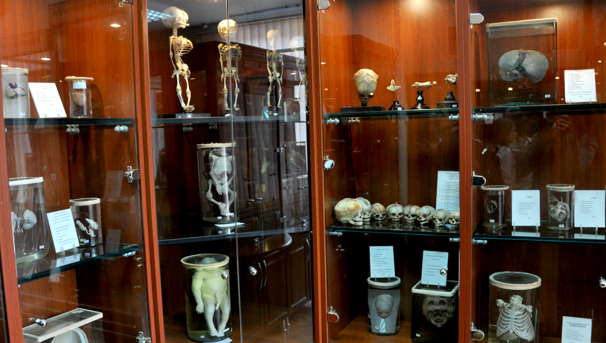
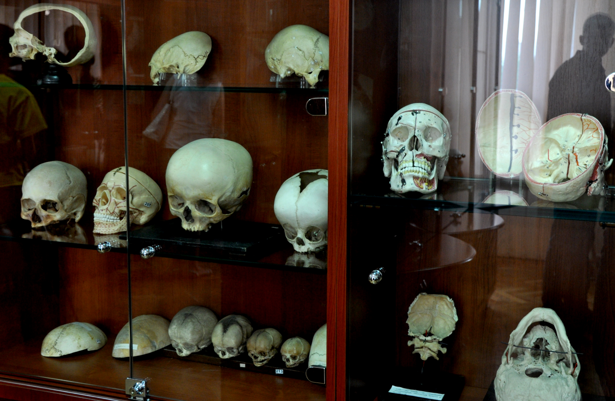
At present, the museum of the Anatomy Department has one of the most valuable and impressive collections of unique anatomic pieces, being among the few of its kind in Europe. It is highly appreciated by international experts. The museum enjoys great popularity, playing an important role in disseminating knowledge about human body, morphological and functional peculiarities at different stages of pre-and postnatal ontogenesis, the influence of different harmful factors and the way of life on the activity of organs and organ systems. It is frequently visited by pupils and teachers from our country’s towns and villages, students from local colleges and universities and many foreign delegations.
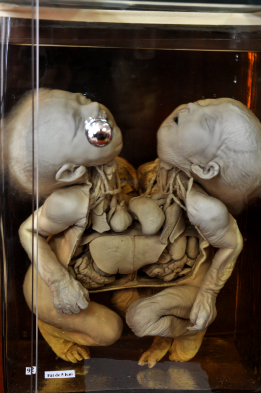
The collection of the Museum of human anatomy includes about 2000 items of high scientific-didactic value, representing bones, skeletons, wet preparations – joints, muscles, internal organs and organ systems, embryos in various periods of development, plastinated anatomical sections, malformations, monstrosities etc., preserved in formalin solution, pieces obtained by corrosion and mummification, anatomical dummies, which constitute a real treasure of our Alma Mater.